The Rivera-Serrano Lab studies how viruses interact with host cells and how those interactions can be harnessed for therapeutic benefit. Our central research program focuses on oncolytic virotherapy: the use of viruses that preferentially infect and kill cancer cells. We use Mammalian Orthoreovirus (family Spinareoviridae), commonly known as reovirus, as both a model system and a potential therapeutic platform because naturally occurring genetic diversity among reovirus strains creates variants with distinct capacities to enter cells, replicate, interact with antiviral defenses, and trigger cancer cell death.
Our work seeks to identify genetically diverse reovirus reassortants with enhanced oncolytic activity against rare cancers whether as monotherapy or in combination with traditional chemotherapeutic approaches. This work provides the foundation for our broader goals: (1) to understand why some virus–cancer cell combinations are more effective than others, (2) identify the host pathways that determine these outcomes, and (3) use that knowledge to improve the selectivity and potency of oncolytic virotherapy.
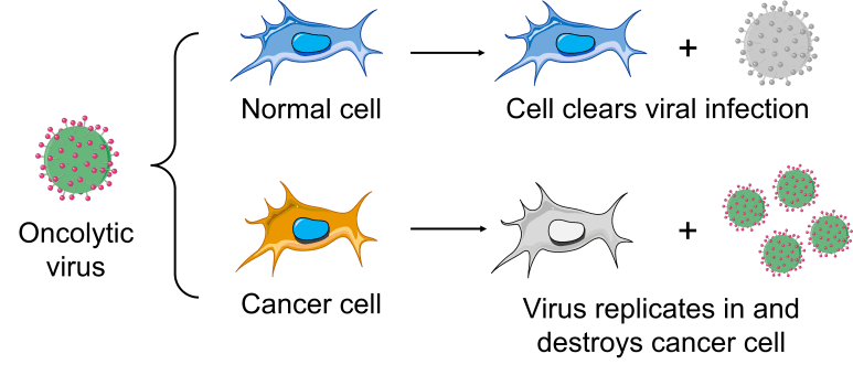
I. Oncolytic virotherapy and virus diversity
Cancer cells often differ from non-transformed cells in antiviral signaling, cell-cycle regulation, stress responses, metabolism, and programmed cell death pathways. These differences can create vulnerabilities that allow some viruses to replicate more efficiently in tumor cells than in healthy tissue. Our lab investigates how genetically diverse reoviruses exploit these vulnerabilities and how specific viral and host-cell factors influence oncolytic potential.
We compare parental reovirus strains, reassortant viruses, and experimentally selected variants across cancer cell models, including fibrosarcoma and epithelial-derived tumor cells. These projects ask:
- Which reovirus variants are most effective at infecting and killing specific cancer cell types?
- Which viral genes or phenotypes are associated with enhanced oncolytic activity?
- How do innate immune signaling and cell-death pathways determine whether infection succeeds or fails?
- Can genetically diverse reoviruses be used to improve therapeutic potential against rare or understudied cancers?
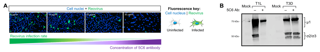
To answer these questions, we combine fluorescence microscopy, cell viability assays, viral protein detection, molecular and cellular biology, bioinformatics, and additional quantitative analyses to connect viral genetics with measurable infection and cancer cell-killing outcomes.
II. Host gene discovery and functional validation of oncolytic pathways
A major goal of our current work is to identify host genes and cellular pathways that determine whether cancer cells are susceptible or resistant to oncolytic reovirus infection. To do this, we are incorporating whole-cell RNA sequencing, or RNA-seq, to compare global gene-expression patterns in cancer cells infected with reovirus variants that differ in their ability to infect, replicate, and kill tumor cells.
These transcriptomic datasets allow us to identify host responses associated with productive infection, antiviral restriction, and virus-induced cancer cell death. We are especially interested in pathways related to innate antiviral immunity, apoptosis, cellular stress responses, viral entry, intracellular trafficking, and cellular metabolism. By comparing susceptible and resistant cancer cell models, we aim to identify host factors that either restrict reovirus replication or make cancer cells more vulnerable to viral oncolysis.
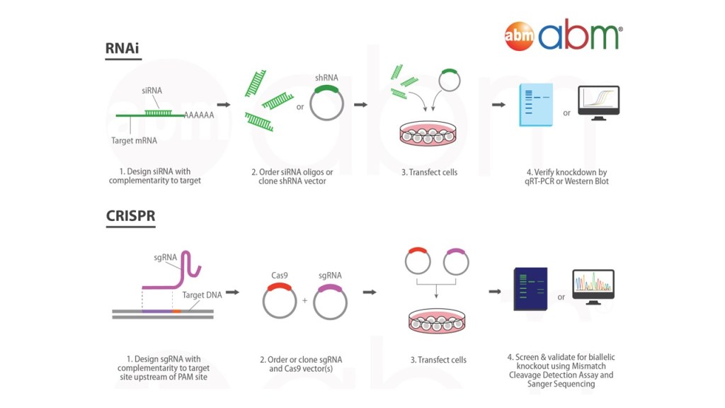
Because RNA-seq identifies candidate genes and pathways, we use functional approaches to determine which of these factors directly control infection outcomes. Using RNAi-mediated knockdown and/or CRISPR/Cas9-based gene editing, we can reduce or eliminate the expression of candidate host genes and test whether those changes alter viral infectivity, viral protein production, antiviral signaling, cancer cell viability, or virus-induced cell death.
Together, these approaches allow us to move from discovery to mechanism. Rather than asking only which genes change during infection, we ask which genes matter, how they influence oncolytic potential, and whether they can be targeted to improve the selectivity and potency of oncolytic virotherapy.
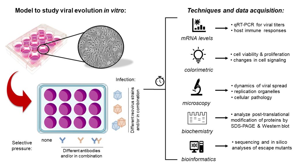
III. Viral evolution and immune evasion
Viruses evolve rapidly, and genetic change can alter how they interact with host cells, immune defenses, and antiviral treatments. Reovirus provides a powerful system for studying viral evolution because its segmented genome allows genetic diversity to arise through both mutation and reassortment, the exchange of genome segments when distinct viruses infect the same cell.
Our lab studies how reovirus evolution shapes virus–host interactions. We are particularly interested in how genetic changes in viral attachment proteins and other viral components influence immune recognition, cell tropism, replication efficiency, and the ability of reovirus to infect and kill specific cell types. These questions are important for understanding viral pathogenesis, immune escape, and the development of improved viral platforms for therapeutic applications.
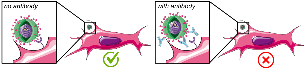
One area of this work uses selective pressure to experimentally direct viral evolution. For example, we infect mammalian cells in the presence of neutralizing antibodies against the reovirus attachment protein σ1. Under these conditions, only viral variants that acquire genetic changes allowing them to escape antibody binding are expected to survive and become dominant over time. These emerging viruses can then be rescued, sequenced, and functionally characterized to determine how specific genetic changes alter viral fitness, immune evasion, and host-cell interactions.
This research connects directly to our oncolytic virotherapy work. By understanding how reovirus evolves to overcome immune barriers, alter cell tropism, and acquire new biological properties, we can better define which viral features contribute to enhanced infection and cancer cell killing. Ultimately, this work helps us understand both the risks and opportunities associated with viral evolution.
IV. Mathematical modeling of within-host cardiac viral diseases
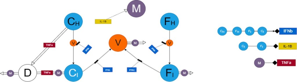
In close collaboration with Dr. Hwayeon Ryu in Elon’s Department of Mathematics and Statistics, our lab also develops interdisciplinary projects that use mathematical modeling to understand how infection, inflammation, and tissue damage unfold over time within the host. These projects combine biological mechanisms with systems-level modeling to identify which cellular interactions or immune pathways most strongly influence disease progression.
This collaboration is exemplified by the work of two recent Lumen Scholars—Elise Butterbach and Lisa Kranec— which models viral myocarditis and investigates how inflammatory and repair pathways can damage cardiac tissue while still helping the host control infection. These projects are particularly suitable for students with a strong background and interest in mathematics, computer science, and engineering.
Through these projects, students learn to move between biology, mathematics, and engineering while asking questions that cannot be fully answered by any one discipline alone. Our long-term goal is to use mathematical models to generate experimentally testable predictions about infectious disease, inflammation, and tissue remodeling.
